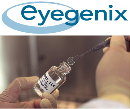
Eyegenix LLC
Overview
I worked for Eyegenix from 2015 to 2018 as a researcher. Eyegenix was a biotech start up company located in Honolulu Hawaii that developed bio-engineered corneas (BEC). We often collaborated and sub-contracted efforts out to those at Johns Hopkins Wilmer Eye Institute. Together with parallel efforts we developed BECs that were sucessful in live transplant. I held two roles while working at Eyegenix. I was a part of the manufacturing team and I was incharge of running biocompatibility assays on developed BECs.
 |
Manufacturing
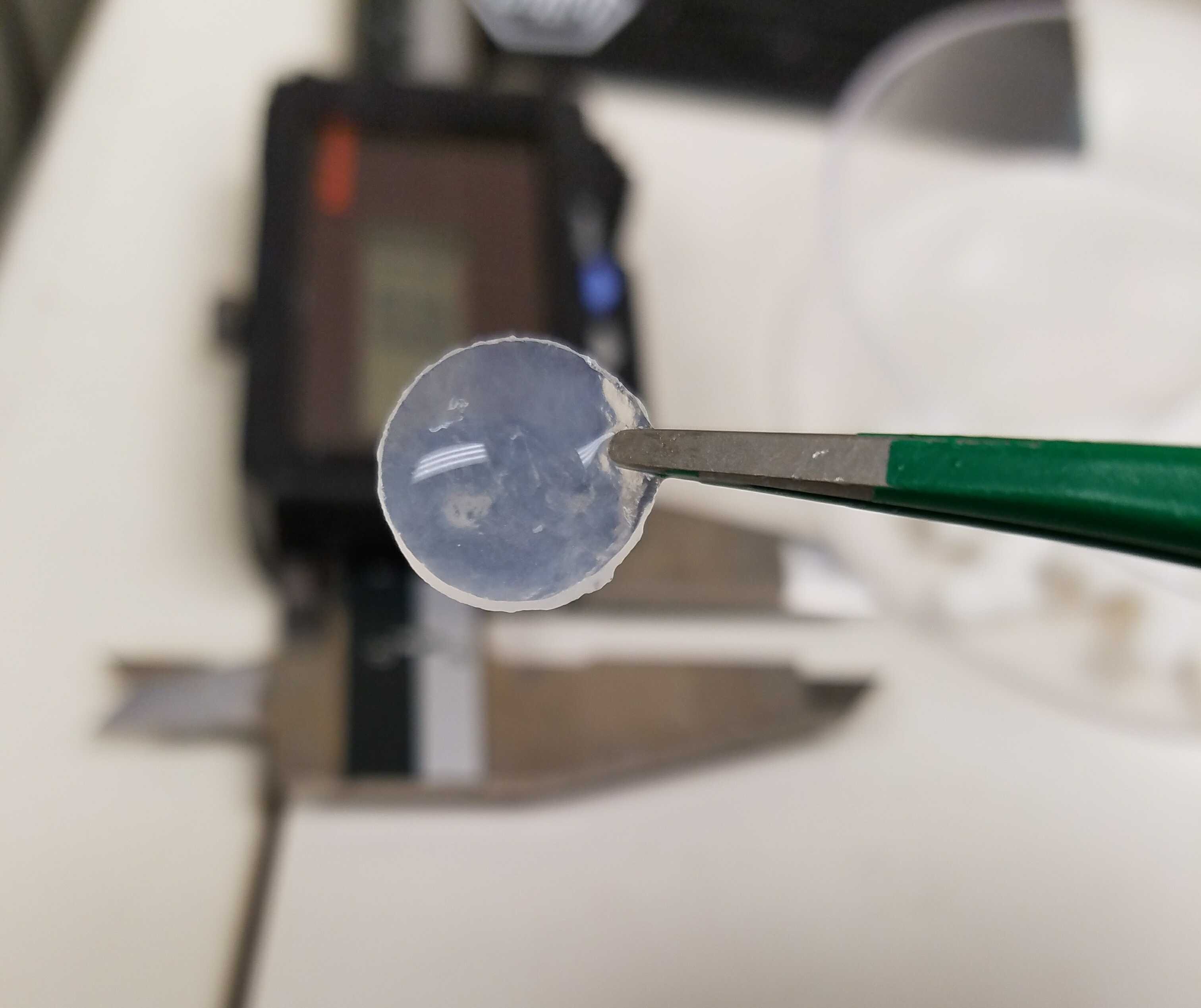
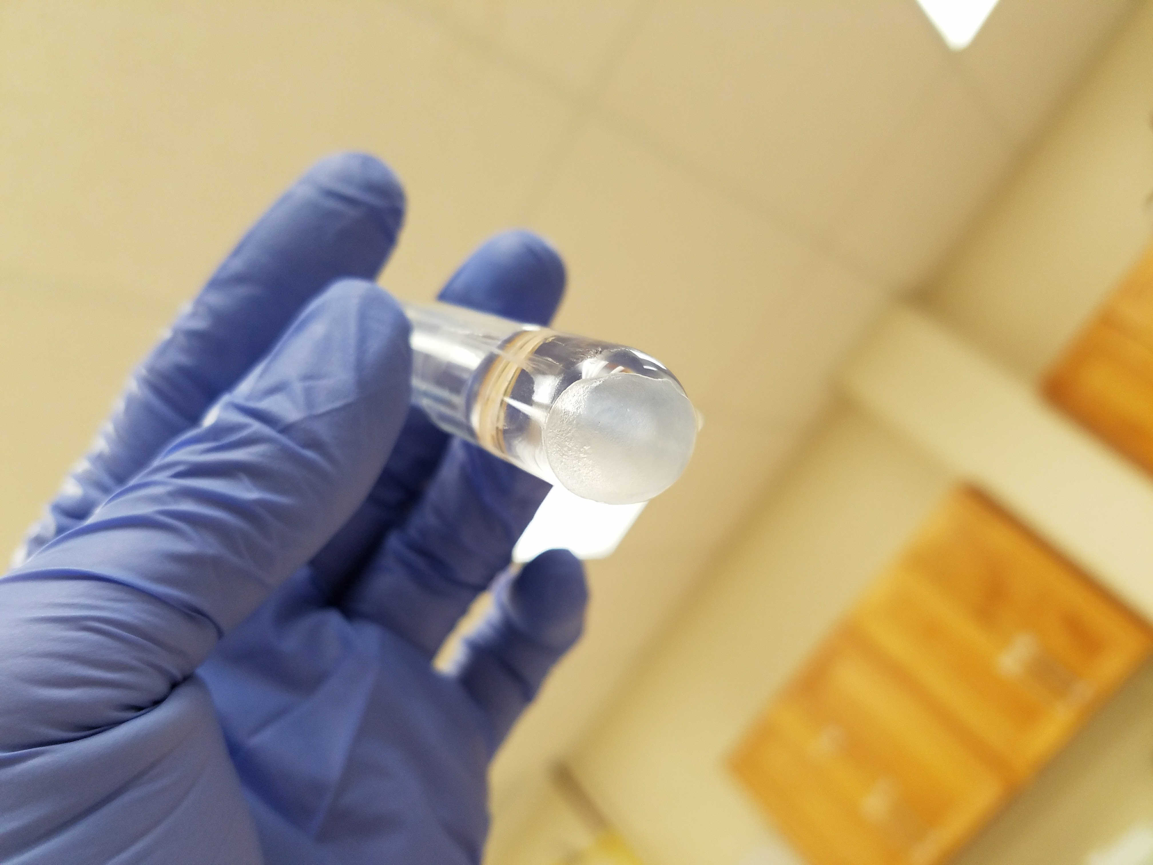
The BEC cosisted of a protein polymer base and units were shaped using injection molding. Eyegenix was in posession of one of two clean rooms on the island of Oahu and this is where units were manufatured.Manufacturing required great dexterity along with familiarity with the feel and viscosity of the polymer during the cross linking stage. As a result, units were manufactured by hand and attempts to automate presented more problems than solutions. As a part of the manufaturing team, I was granted the privilege of working with the polymer and building units for transplant. Additionally, I participated in the development of new polymer formulations. New polymers were formulated with a goal to improve strength, elasticity, biocompatibility, and clarity. Deriving new formulations allowed me to exercise my biochemistry knowledge in a way that produced tangible and testible results.
 |
Biocompatibility Testing
I was originally hired to performe cell and tissue culture task. Test were performed in a bio-safety level 2 rated lab using imortalized human and primary rabbit cornea epithelial cells. Two biocompatibility assays were develped to assess how BECs will perform in-vivo.
The first test was called a direct contact assay which tested the accute bio-compatibility of our BEC. Cells were placed on the BEC and left to grow for a seven days. After 7 days, the living cells were quantified using spectoscopy and MTT. The second assay was the cell migration assay which evaluated how well BEC materials promote cell migration to cover the material. An area in the middle of the test BEC was occluded and cells were places on the covered portion of the bec. Image analysis with the NIH’s imageJ was used to quantify migration rates over the BEC surface. Images were taken under a microscope at periodic time points.
 |
While working in tissues culture, I was able to practice and improve my wet lab skills. Contamination is always a conern when performing tissue culture and I was able to perfect my sterile technique. I also learned to maintain a proper inventory as well as a clean and sterile work space.